
Breast Ultrasound
Staff Contact
Maribeth Girard
Certification Specialist

Breast Ultrasound Certification
Sample Test Questions
Category: Anatomy
The hilum of a normal lymph node is usually:
- Hypoechoic
- Hyperechoic
- Isoechoic
- Not visible sonographically
Category: Physics
Focusing:
- Improves lateral resolution
- Improves axial resolution
- Increases beam width in the focal region
- Shortens pulse length
Category: Scanning Techniques
When scanning the breast, the transducer may be moved in a direction parallel to the long axis of the foot plate. This "skiing" motion will cause the image of a lesion visualized in the breast to:
- Appear and disappear at a given point on the screen
- Move from left to right or right to left across the screen at a constant depth
- Appear at a deep position on the screen and disappear at a superficial position on the screen
- Move diagonally across the screen
Category: Anatomy
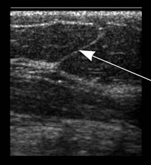
In this breast ultrasound image, the arrow indicates:
- Cooper's ligament
- Breast parenchyma
- A vessel or dilated lymphatic
- Scanning artifact